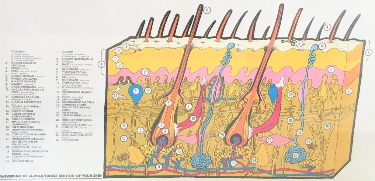
The anatomy of the pilosebaceous follicle
The number of pilosebaceous follicles on the whole body is estimated at 5 million.
The number of hair follicles on the cephalic end is estimated at 1 million.
Each hair and hair consists of an extra-cutaneous part or hair proper and an intra-cutaneous part called the pilosebaceous follicle burrowing up to 3 to 4 mm below the surface of the skin.
The pilosebaceous follicle is a tubular invagination of the epidermis in which the hair itself is lodged.
One two or three pilosebaceous follicles together with the sebaceous gland and the pilosebaceous muscle form an entity called the pilosebaceous follicular unit.
The sebaceous gland derives from a bud of the external epithelial sheath GEE (itself in continuity with the epidermis).
The sebaceous glands are located inside the connective tissue sheath surrounding the follicle.
The density of sebaceous glands is 100 / cm2 up to 400 at the level of the front face of the scalp.
Sebum, the secretion product of the sebaceous gland, is made up of lipids and cellular debris. The piloerector muscle is inserted on the fibrous or conjunctive sheath of the hair.
At the level of the extra-cutaneous portion, that is to say after the hair has come out of the infundibular ostium, the hair is alone: the hair shaft or the extra-cutaneous part of the hair itself.
The intra-cutaneous portion or also called the root comprises the pilosebaceous follicle and the hair itself.
The upper part of the pilosebaceous follicle, ie the area from the surface of the skin or ostium to the insertion zone of the pilosebaceous muscle, is the permanent zone of the pilosebaceous follicle.
It remains present throughout the hair cycle and consists of:
-The infudibulum: this area extends from the surface of the skin or ostium to the mouth of the sebaceous gland or collar.
The infundibulum does not vary during the cycle.
- The multiristified epithelium: keratinized which borders the infundibular canal is continuous with the surface epithelium or epidermis.
-The isthmus: is the area which extends from the area where the sebaceous gland ends up to the insertion of the pilo-erector muscle. The isthmus does not vary during the cycle.
- The bulge: is a bulge of the external epithelial sheath, located at the level of the insertion of the pilo-erector muscle at the union of the middle 1/3-1/3 upper of the pilosebaceous follicle. This zone is very well individualized morphologically in mice.
This area contains cells with a high germination potential, which are the source of the re-epithelization of the epidermis. These cells have been identified by immunochemical studies using stem cell markers (CD 34, keratin 15, keratin 19).
The bulge area is less well individualized in humans than in other species.
The lower part of the pilosebaceous follicle extends from the muscle insertion to the base of the bulb. It is the temporary part of the follicle or also called the area of the epithelial sheaths. This portion varies with the different phases of the hair cycle.
She understands
-A first portion, called by some region the lower hair shaft, extends from the insertion zone of the piloerector muscle to the Adamson fringe.
- The Adamson fringe is the region of transition between keratinocytes in the process of keratinization still alive and completely keratinized dead cells.
-The deepest part of the pilosebaceous follicle is formed by the bulb and the follicular or dermal papilla.
The bulb is the region from the Adamson fringe to the base of the follicle.
The bulb includes
-The pigmented area: the area where the hairs are loaded with melanin.
-The keratogenesis zone: which is more superficial and which corresponds to the keratinization zone. In the keratinization zone, the diameter of the hair decreases significantly due to the dehydration of the keratinocytes.
-The germinal zone: zone of proliferation of keratinocytes.
The hair matrix is the deep area of the pilosebaceous follicle.
The hair matrix is the growth area of the pilosebaceous follicle.
At the level of the matrix, the epithelial cells multiply and migrate upwards giving rise to the cells of the hair shaft (medulla, cortex and inner cuticle) and cells of the internal epithelial sheath GEI (outer cuticle, Huxley's layer and Henle).
The matrix is the widest part of the hair.
The area of the matrix containing the melanocytes is pigmented.
Above, the area containing cells not yet keratinized is clear
The area of the start of keratinization is darker.
As previously described, the pilosebaceous follicle is partly composed of epithelial elements and partly dermal elements.
Thus the hair shaft or hair itself is made of three portions.
- the medulla: chestnut color is made of a layer of clear bulky keratinocytes, sometimes pigmented, little keratinized comparable to the keratinocytes of the stratum spinosum. The medulla is absent in case of down and lanugo
Distally, the keratinocytes of the medulla are transformed into schizokeratin.
-the cortical, cortex, bark: represents 90% of the weight of the hair. The cortex is made up of several layers of ill-defined oval keratinocytes elongated in the direction of the axis of the hair loaded with melanin more or less pigmented depending on the color of the hair, firmly attached to each other and elongated in the direction of the hair. The keratinocytes of the cortex fill from the bottom up with keratin through a process of keratinization. Higher up the keratinocytes of the cortex are transformed into sclerokeratin.
the internal cuticle or internal epirermicle is formed of a keratinocyte layer welded to each other by proteins and ceramides. The keratinocytes are stacked on top of each other. Their free edge is directed towards the distal end of the hair.
Hair pigmentation
As a reminder, there are two types of melanin: Eu-melanin (brown) and Pheo-melanin: (yellow red), the synthesis of which is made from tyrosine via DOPA and DOPAquinone.
The pigment load produced by the pigmentation unit, therefore the color of the hair, depends on the phototype, on the phase of the hair cycle.
Active melanocytes are located at the apex of the papilla and constitute the unit of pigmentation which is destroyed during the anagen-catagen transition. These melanocytes, which are very numerous at the bulb level, distribute the melanin produced to the bulbar keratinocytes which will then keep this melanic content throughout the differentiation process.
Melanocyte activity is cycle dependent.
The production of melanin is very important during the late anagen phase.
At the very beginning of the anagen phase, the melanin load is low.
Melanogenesis stops during the catagen phase.
The activity of melanocytes stops before that of keratinocytes during the catagen phase. Thus the intra-cutaneous part of the hair shaft during the catagen and telogen phase is weakly pigmented
Melanin is present in the hair shaft and in the bulb.
The concentration of melanin would be higher at the level of the medulla than at the level of the cortex.
The GEI is not pigmented, the GEE would not be rich in melanin despite the presence of melanocytes at its level. These GEE melanocytes participate only slightly in hair pigmentation.
Keratin
Hair, a natural fiber, is made up of 95% keratin, a hard and fibrous protein coming from dead cells which is also the main constituent of our nails. Keratin is a scleroprotein rich in sulfur (ie a protein made up of a large number of amino acids), often seen in the form of plaques arranged in tiles. This is insoluble in water and ensures impermeability and protection against external agents acting on the hair. The keratin fibers are tied together for more strength, by bonds of different types, the most important of which are those in which there is sulfur. Keratin is produced by keratinocytes, which have a lifespan of thirty-five to forty days.


